What are the signs of metabolic bone disease (MBD) in reptiles?
Short answer
Early MBD shows as soft or rubbery jaw, tremors in the limbs, slow or twisted growth, and weak hind legs. Advanced MBD shows as bone deformity (kinked spine, bent limbs), pathological fractures, seizures and inability to walk. MBD is caused by chronic low UVB, low dietary calcium, or excess phosphorus — and is almost entirely preventable with correct UVB and a Ca:P 2:1 diet.
- Author
- Reptimo Editorial
- Updated
- Updated
- Reading time
- 6 min read
What MBD actually is
Metabolic bone disease is the umbrella name for a group of disorders in reptiles where calcium homeostasis fails. The most common form in captive reptiles is nutritional secondary hyperparathyroidism: blood calcium drops, the parathyroid glands respond by pulling calcium out of bone to maintain it, and over weeks-to-months the skeleton progressively demineralises. The PetMD MBD reference identifies this as the most common preventable health problem in captive reptiles.
The clinical end-points are visible — rubber jaw, kinked spine, bent limbs, fractures — but the disease process is silent for months before those appear. Which is why the framework matters more than the symptom list: catch the deficiency upstream and the bone never demineralises in the first place.
Why MBD happens — the three legs
Three causes, almost always combined:
Care parameters
The three causes of MBD in captive reptiles
| Parameter | Recommended value | Notes |
|---|---|---|
| Insufficient UVB | Stops vitamin D3 synthesis | Without D3, gut can't absorb dietary calcium |
| Low dietary calcium | Un-dusted, un-gut-loaded prey | Common when feeding insects without supplementation |
| Excess dietary phosphorus | Binds calcium, makes it unavailable | All-mealworm or all-fruit diets are classic risks |
Any single leg failing pulls calcium homeostasis over months. All three failing — a juvenile bearded dragon on a coiled bulb, un-dusted crickets and lettuce — produces MBD in 8–16 weeks.
Who gets MBD
Risk is dramatically species-dependent:
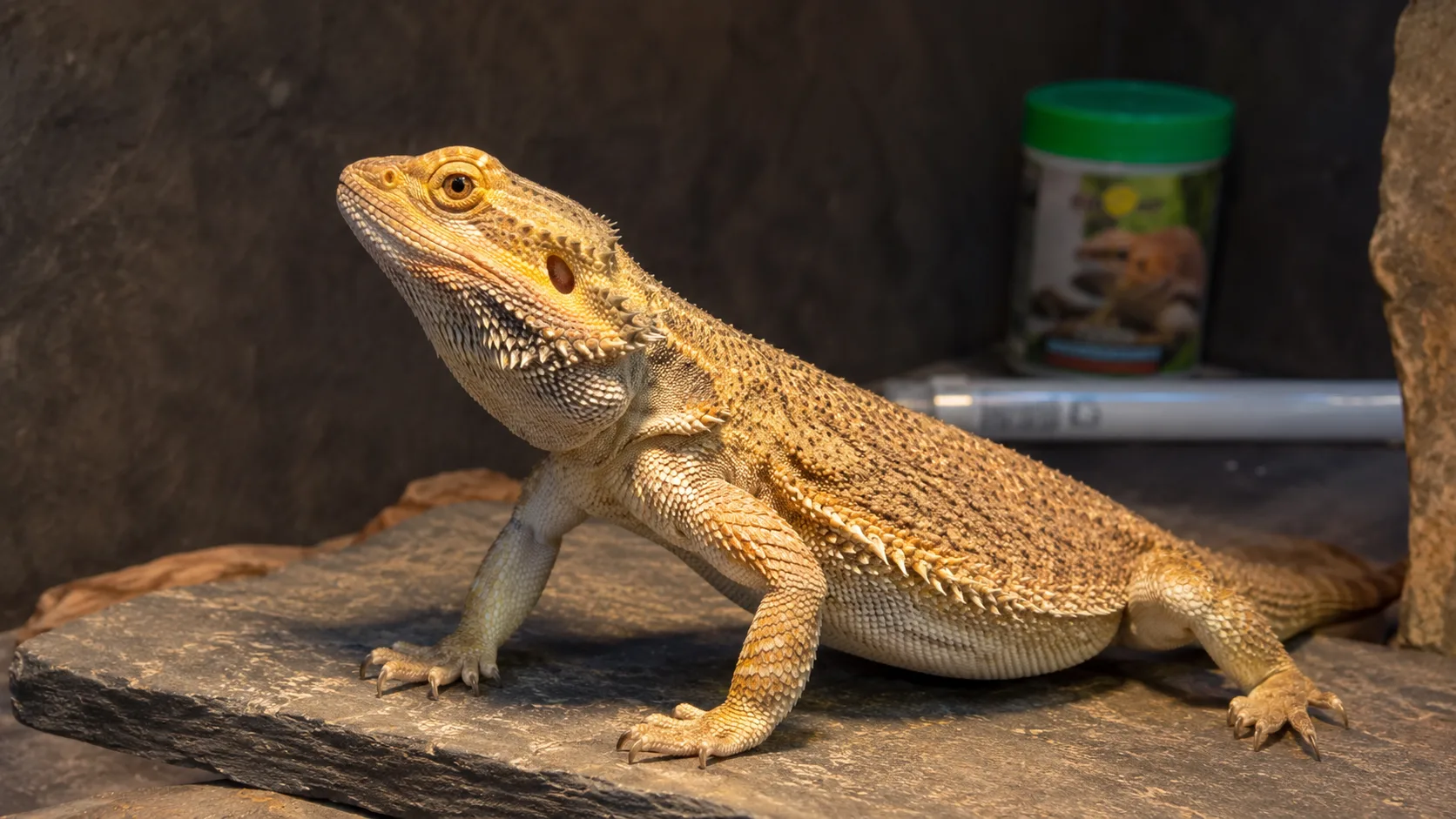
- Highest risk: diurnal heliothermic baskers — bearded dragons, uromastyx, sliders, tegus, savannah monitors, panther chameleons, day geckos. These evolved on strong UVB and high dietary calcium.
- Moderate risk: nocturnal and crepuscular species with low UVB needs — leopard geckos, crested geckos. Still vulnerable on a chronically low-calcium diet.
- Lower risk: snakes fed appropriate whole rodents. Whole prey delivers calcium and D3 with minimal supplementation needed. Risk rises with poor-condition prey or insect-only diets (rare).
For species-specific context, see bearded dragon MBD signs and the cross-species UVB guide.
Early signs
The early signs are what to catch — by the time the late signs appear, bone is already deformed:
- Rubbery or soft jaw on palpation. The lower jaw feels pliable rather than firm. Best detected by a vet or experienced keeper.
- Fine tremors in the limbs, often after activity or feeding.
- Hind-leg weakness or dragging, intermittent.
- Twisted or wide-stance gait.
- Slowed or paused growth in juveniles. Often the very first sign in fast-growing species.
- Reluctance to bask or climb.
A reptile showing any one of these signs in combination with a known husbandry gap (old UVB tube, no calcium dusting, poor diet) goes to a vet. Don't wait for kinking or fractures.
Advanced signs
These are obvious — and indicate weeks-to-months of established disease:
- Visible bone deformity — kinked spine, bent limbs, shortened or misaligned lower jaw ("rubber jaw"), swollen joints.
- Pathological fractures from minor handling.
- Seizures and twitching, especially in advanced hypocalcaemia.
- Complete inability to walk or right itself.
- Constipation and bloating from intestinal smooth-muscle weakness.
Per the Merck Veterinary Manual, seizures from hypocalcaemia in advanced MBD are a medical emergency — vet within hours, not days. The same source notes that bone deformities already in place are usually permanent even after treatment.
How a vet confirms MBD
A reptile-experienced vet uses three tools:
- X-ray — shows bone demineralisation, fractures, deformity. The classic finding is "pencil-thin" cortical bone.
- Blood panel — calcium, phosphorus, ionised calcium, and parathyroid hormone where available. Calcium-to-phosphorus ratio inversion is diagnostic.
- Physical exam — palpation of jaw and limbs, body condition, gait observation, response to handling.
Find a vet through the Association of Reptilian and Amphibian Veterinarians (ARAV) directory — most general small-animal vets see few reptiles and may miss early MBD.
Treatment
Treatment depends on severity but always combines husbandry correction with medical support:
- Husbandry correction — install a fresh species-appropriate T5 HO UVB tube at the correct distance, fix the diet and supplementation protocol immediately. This is non-negotiable; no treatment works without it.
- Calcium gluconate injections (vet-administered) for acute hypocalcaemia.
- Oral calcium glubionate for ongoing supplementation, dosed by the vet based on weight.
- Vitamin D3 injection in selected acute cases — used carefully, as D3 overdose is harmful.
- Supportive care — assist-feeding if not eating, fluid therapy if dehydrated, cage rest if fracture risk.
Recovery takes weeks to months. The keeper-side job is consistent husbandry and supplementation through that window.
How to prevent MBD
Prevention is genuinely straightforward — three habits, sustained:
- Correct UVB for the species.
- Diurnal baskers (bearded dragons, sliders, uromastyx) — T5 HO 10.0/12 % at the right distance, UVI 4–6 at basking, replaced at 12 months.
- Low-UVI species (leopard geckos, crested geckos) — T5 HO 5.0 / 7 %, UVI 0.5–1.5 at basking.
- See the cross-species UVB guide for the full Ferguson-zone table.
- Calcium-with-D3 dusting on insects or greens:
- Juveniles: 3–5 times a week
- Adults: 2–3 times a week
- Diet with Ca:P ~ 2:1 — gut-load insects on calcium-rich greens, avoid all-mealworm or all-spinach diets that load phosphorus or bind calcium with oxalates. Include a multivitamin every 1–2 weeks.
When MBD becomes a vet emergency
Some signs move MBD into an immediate vet category:
- Seizures or twitching from hypocalcaemia.
- Inability to walk, lift the head, or right when flipped.
- Pathological fracture from minor handling.
- Marked bloating with constipation.
For the broader cross-species warning-signs framework that MBD signs sit within, see "is my reptile sick?".
After diagnosis
A reptile recovering from MBD needs the husbandry and supplementation protocol logged and sustained for life — not just until symptoms improve. The conditions that caused it will cause it again if relaxed. A tracking app that reminds about UVB replacement and supplementation cadence is genuinely useful here; for the format options, see the tracking-format comparison.
Frequently asked questions
What is metabolic bone disease in reptiles?
What causes MBD in pet reptiles?
What are the earliest signs of MBD?
Can MBD be reversed?
Which reptiles get MBD most often?
How do I prevent MBD?
Can a snake get MBD without UVB?
What does the vet do for MBD?
Is rubber jaw always MBD?
Sources
- Metabolic Bone Disease (MBD) in Reptiles · PetMD
- Disorders and Diseases of Reptiles · Merck Veterinary Manual
- What Are Ferguson Zones? · Zen Habitats
Quick check
Test what you just learned
A short quiz, just for you. Pick an answer to get instant feedback — there's no pass mark, this is for your benefit.
Quiz questions and answers
Which of these is the earliest sign of metabolic bone disease in a reptile?
Correct answer: A soft or rubbery jaw on palpation, plus subtle tremors after exertion
Subtle signs — soft jaw, fine tremors, slightly twisted gait, hind-leg weakness — come weeks to months before visible bone deformity. By the time the spine is kinked or a fracture appears, MBD is well-established. Early signs are the ones to catch.
What's the three-part recipe that prevents MBD in a captive reptile?
Correct answer: Correct UVB for the species + calcium-with-D3 dusting + gut-loaded insects on a Ca:P 2:1 diet
Prevention is a three-legged stool: appropriate UVB (T5 HO tube at the right Ferguson-zone UVI, replaced at 12 months), regular calcium-with-D3 dusting, and a diet with a Ca:P ratio of around 2:1. Drop any leg and MBD risk climbs over months.
Why do diurnal baskers (bearded dragons, sliders, uromastyx) get MBD more often than snakes?
Correct answer: Diurnal baskers evolved on strong UVB and high dietary calcium — without both in captivity, deficits accumulate fast; snakes get most calcium from whole prey
Diurnal heliothermic baskers depend on UV-driven D3 and a Ca-rich diet. Snakes get most calcium and D3 from whole prey (rodents) and were thought not to need UVB. Skip UVB for a bearded dragon and MBD develops within months; skip UVB for a well-fed ball python and risk is low.